Circulation
Let’s go back to blood circulation.
The uterus with the baby and other contents weighs approximately five kilos.
What happens?
The blood comes from the inferior limbs and that flows into the cava vein has its flow diminished in this region, delayed at the legs. In consequence a lesser volume of blood arrives at the heart. The organism receive that message and turns on a compensatory mechanism to protect the heart and the brain.
The vessels that take blood to the kidneys and to the uterus are contracted, to keep up the volume of blood in circulation. Those are the organs of liquid elimination.
And who is inside the uterus? The baby.
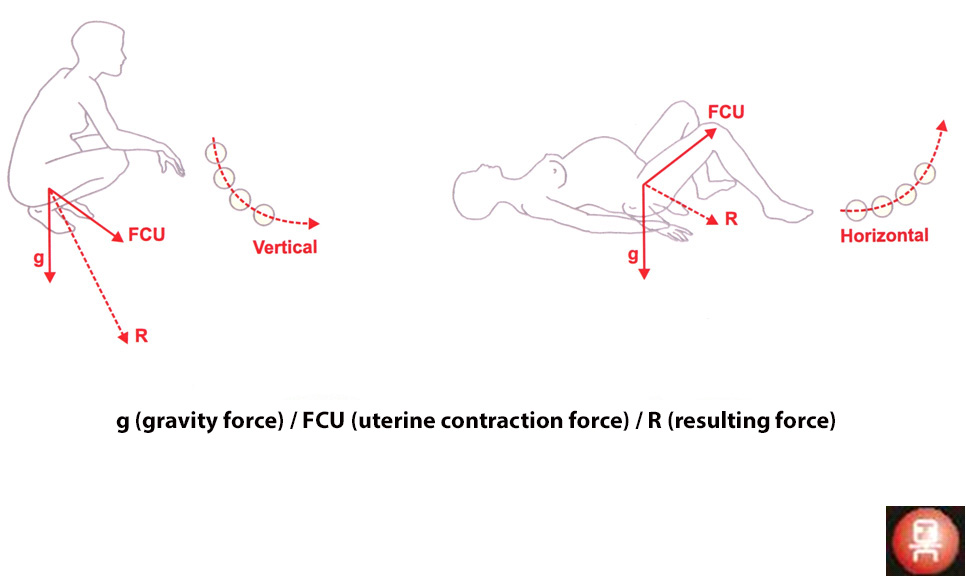
When the uterus drops over the cava, the baby receives less oxygen, directly, by the compression over the aorta and mainly by the consequences of the compression of the cava vein. The blood that comes from the uterus in the direction of the cava vein has a higher pressure coming back, which is transmitted to the placenta, facilitating the premature loosenings. That mechanism happens more frequently in older women with more children, varicose veins and diminished muscular tone.
In 10% of pregnant women the blood pressure falls because of that, arriving even at extreme cases of shock from supine hypertension. That happens in all epidural anestesias, and the anesthesists push the uterus to the left to avoid the drop in blood pressure.
We have measured the circumference of the abdômen in 200 women on the ninth month of pregnancy standing up, lying down and squatting.
The cincumference was smaller by 4 cm when the future mother was lying down, showing the approach of the uterus to the cava vein.
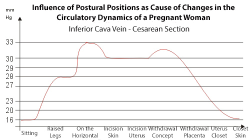
One of the experiments we have performed to verify the influence of the pregnant women’s position on the blood flow dinamics consisted in the installation of a cateter, linked to a manometer – “pressure vein” – putting in on the cava veins and on the right atrium.

It was done with a pregnant patient, suffering of cardiac insufficiency, who was going to be submitted to a cesarian operation. The blood pressure was taken lying down, squatting, sitting down and after the epidural anesthesia, during the surgery.
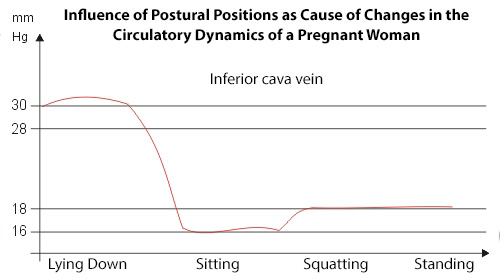
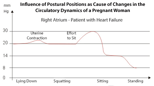
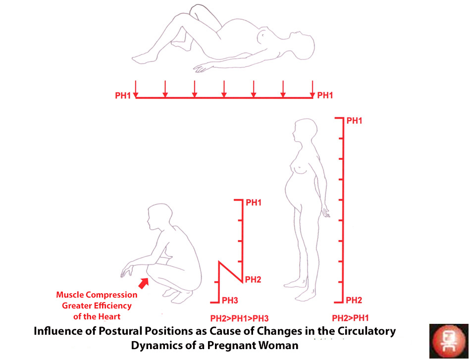
The higher the pressure, the less blood flows. The pressure gets higher with the effort when the uterine contraction takes place. On the inferior cava vein the pressure is higher when the patient is lying down than in other positions. When the epidural anesthesia was perfomed, the pressure more than doubled and only came back to the levels of sitting down position after the placenta was taken out.
The best position is squatting, with no effort. The flexed legs press the veins of the inferior limbs, in the direction of the right atrium.
The hidrostatic pressure is higher between the navel and the pubis, where the uterus is situated, allowing a better irrigation of that region with consequently better oxigen flow.
The birth channel in the horizontal position shows its narrowing, since the sacrus bone cannot move backwards, because it lies over a hard surface, the bed.
The opposite happens when the person squats, all the pelvic bones can expand with no obstacles.